Bone Growth Stimulation for Nonunion Fractures
 How Does a Bone Growth Stimulator Work?
Bone growth stimulation is a form of treatment that is used to promote the healing of nonunion fractures, which refers to a bone fracture that does not display any visible signs of healing [1]. The stimulator is a small device that can be implanted under the skin or outside of the skin and it administers small electrical pulses to the area of the fracture in order to promote bone growth and healing. The device is easily concealed under the clothes and does not cause painful, electrical sensations.
Bones are made up of living tissue and a fracture results in two broken ends; one of which has a positive charge and the other end which has a negative charge. The stimulator generates an electromagnetic field that helps attract the two oppositely charged ends of the bones to each other by promoting new bone growth and fusion, which leads to healing. There are four main stages of fracture healing: inflammation, soft callus formation, hard callus formation, and bone remodeling [2].
Inflammation occurs immediately after the bone is broken. It develops due to the disruption of the blood supply and bleeding from the fractured ends, which causes swelling and bruising at the fracture site. Soft callus formation refers to the formation of cartilage between the two fractured ends, and this begins a couple of days after the fracture has occurred. Hard callus formation begins around 2 to 3 weeks after the fracture and is the stage when the cartilage starts to transform into bone. This stage continues for 6 to 12 weeks. Bone remodeling begins after the fractured ends have fused together and refers to the formation of extra bone at the site. Unfortunately, in the case of nonunion fractures, this process does not take place as it normally would [1, 2].
Non-union Jones fractureThere are several reasons why fractures may not heal properly on their own, such as: a large gap between fractured bones, improper immobilization of the fractured bone, an infection, improperly aligned fracture ends, or inadequate vascular structures within the fracture site [1]. Bone growth stimulators promote the healing of fractured bones by stimulating the growth of new bone which begins to fuse the broken ends together. If successful, the treatment will help the bone regain its form and mobility. There are several different types of bone growth stimulators, but electromagnetic and ultrasound stimulators are currently the most commonly used devices [1].
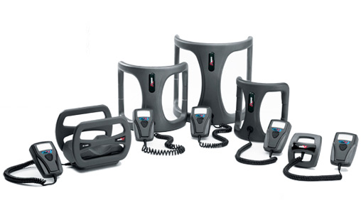
Types of Stimulators
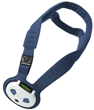
Cervical bone growth stimulator
A direct current (DC) stimulator was one of the first types of stimulators that was used to promote the healing of nonunion fractures. It involves surgically implanting electrical wires of different lengths directly into the fracture site, as along with a lithium battery [3, 4]. There are several disadvantages of DC stimulators which include: the battery life is only about 6 to 8 months, it is hard to properly place the wires and battery under the skin, the stimulator may short circuit if the wires come in contact with each other, a risk of infection, and a second procedure is necessary to remove the hardware [5]. Electromagnetic and ultrasound stimulators gained popularity shortly after the introduction of DC stimulators.
Electromagnetic bone growth stimulators, which are most commonly used, are external devices that have been divided into two categories: capacitive coupling or inductive coupling devices. Capacitive coupling (CC) devices may be in the form of electrodes or pads placed on the skin, at the fracture site, that produce an electrical field of 1-100 mV, and an external battery pack that supplies the electrodes with 20-200 kHz [4]. The stimulator is lightweight and small, but the electrodes may cause skin irritation depending on how often the stimulator is used [6].
Inductive coupling is also referred to as pulsed electromagnetic fields (PEMFs) and this type of bone growth stimulator is made up of a coil that is sized to fit over the fracture site, but it is typically placed under a cast or inside a brace so that it comes in direct contact with the skin. An external battery supplies the PEMFs, which creates an electrical current in the nonunion fracture that promotes healing [1, 4]. The PEMF stimulator tends to be heavier than the CC stimulator and this causes some people to stop using it.
A combined magnetic field stimulator (CMF) may also be used for nonunion fracture treatment and this device combines a DC electrical field with current produced from an external coil. This stimulator is typically worn for 30 minutes daily, which often improves patient compliance for an external device. CMF has been shown to effectively stimulate new bone growth and fracture healing [1, 6].

CMF bone growth stimulator
The average range is between 30 minutes to 9 hours a day, depending on the brand. For longer treatment times, the treatment can be split up into several shorter sessions throughout the day. The treatment period may be anywhere from 3 to 12 months [4]. The use of a stimulator may be contraindicative for pregnant women as well as individuals who have defibrillators or pacemakers. In addition, in order to avoid bone degradation, the current density should not be higher than 625 mA/in2 [1].
Another type of bone stimulator is the low-intensity pulsed ultrasound (LIPUS), and it involves the use of sound waves that are transmitted through both soft tissue and bone. The waves result in micro-motion or mechanical stress at the fracture site, which stimulates the increased production of various cellular components (e.g., proteins, enzymes) and bone growth that initiates fracture healing [2 , 7]. The stimulator is most effective when it is used for 20 minutes a day at specific parameters that have been shown to increase essential nutrients, blood flow, and bone tissue growth at the fracture site [7]. Fracture healing from a LIPUS stimulator may begin anywhere from 2 weeks to 2.5 months after treatment has started [2, 8].
Diagnosis for Treatment
Lateral malleolar fractures (a broken ankle), radial fractures (a broken wrist), and tibial fractures (a broken shinbone) are commonly treated with bone growth stimulation if the following criteria are met [2, 9]: 1) radiographic evidence has confirmed that nonunion long bone fracture healing has ceased for 3 months or more, 2) nonunion of long bone fracture has been documented by a minimum of two sets of radiographs separated by a minimum of 90 days, which demonstrate multiple views of the fracture site along with a written explanation from a clinician stating that no clinically significant evidence of fracture healing has been observed between the two sets of radiographs.
Bone growth stimulation is also used to help improve the outcomes of spinal fusion surgery, which is a procedure that involves fusing two or more spinal bones (vertebrae) together in order to restore the stability of the spine [10]. Spinal fusion surgery entails removing pieces of bone from another part of the body, often the hip, and using the segments to make a type of bridge between the spinal bones that need to be fused together. After the surgery, new bone growth usually takes places over several months and even up to one year. If the procedure is successful, the grafted pieces fuse into solid bone. The use of a bone growth stimulator enhances the healing process, especially in patients who have an increased risk of suffering from a failed fusion. For example, individuals who are undergoing a multi-level spinal fusion are susceptible to healing problems. PEMS bone growth stimulation, in particular, has demonstrated positive results for patients undergoing spinal fusion surgery [10].
Treatment success rates are fairly stable among the various forms of bone growth stimulators and the occurrence of potential side effects depends on the health of the patient, the type of stimulator that is used, and whether stimulation is administered properly [1]. The improper delivery of the electromagnetic or ultrasound stimulation may cause bone tissue damage or degradation. Furthermore, the implantation of internal devices carry more risks (e.g., infection) than external devices. To date, bone growth stimulators are demonstrating increasing success in treating nonunion fractures, especially following failed reconstructive surgeries.
References
- Behrens SB, Deren ME, Monchik KO. A review of bone growth stimulation for fracture treatment. Curr Orthop Pract. 2013; 24(1):84-91.
- Mundi R, Petis S, Kaloty R, Shetty V, Bhandari M. Low-intensity pulsed ultrasound: Fracture healing. Indian J Orthop. 2009 Apr;43(2):132-40.
- Black J. Electrical Stimulation: its Role in Growth, Repair, and Remodeling of the Musculoskeletal System. New York: Praeger; 1987.
- Aaron RK, Ciombor DM, Simon BJ. Treatment of nonunions with electric and electromagnetic fields. Clin Orthop Relat Res. 2004; 419:21–29.
- Paterson DC, Lewis GN, Cass CA. Treatment of delayed union and nonunion with an implanted direct current stimulator. Clin Orthop Relat Res. 1980; 148:117–128.
- Nelson FR, Brighton CT, Ryaby J, et al. Use of physical forces in bone healing. J Am Acad Orthop Surg. 2003; 11:344–354.
- Claes L, Willie B. The enhancement of bone regeneration by ultrasound. Prog Biophys Mol Biol. 2007 Jan-Apr; 93(1-3):384-98.
- Victoria G, Petrisor B, Drew B, Dick D. Bone stimulation for fracture healing: What’s all the fuss? Indian J Orthop. 2009 Apr;43(2):117-20.
- Medicare claims processing manual. In: Services CFMaM, ed. Billing Requirements for Special Services. Baltimore, MD: Department of Health and Human Services, United States Government; 2010.
- Kaiser MG1, Eck JC, Groff MW, Ghogawala Z, Watters WC 3rd, Dailey AT, Resnick DK, Choudhri TF, Sharan A, Wang JC, Dhall SS, Mummaneni PV. Guideline update for the performance of fusion procedures for degenerative disease of the lumbar spine. Part 17: bone growth stimulators as an adjunct for lumbar fusion. J Neurosurg Spine. 2014 Jul;21(1):133-9.