What is a Non-union fracture?
When a fracture site has failed to heal and shows no visibly progressive signs of healing it is established as non union [2].
How is a non-union fracture different then a delayed union fracture?
Delayed union: A fracture that requires more time than usual to heal but shows progression [1].
Nonunion: A fracture that has not healed in an extended period of time and shows no signs of potential for healing.
What are the common symptoms of a non-union fracture?
Symptoms can include swelling, pain, tenderness, deformity, and difficulty bearing weight.
What area of the body is most likely to develop a non-union?
The most common site of non-union fracture is of the tibia, which is the larger bone of the lower leg. Other common sites are the neck of the femur, the large bone connecting the hip joint to the thigh, and smaller bones in the foot and hands.
How are non-union fractures classified?
There are four classifications of non-union fractures
- Hypertrophic- a large lipped region called a callous forms but the bone does not heal. This gives the appearance of an elephant foot on X-Ray. If healing is less aggressive images can resemble a horse’s hoof.
- Oligotrophic- a small callus, to heal the bone, forms, but the healing process is not aggressive even though there is adequate blood supply.
- Atrophic/Avascular-no callus forms and the region is lacking blood and oxygen needed for healing. This is called ischemia.
- Pseudarthrosis- excessive motion and instability creates a false joint in the bone over time.
What are the risk factors for a non-union fracture?
Common risks factors for non-union fractures include: smoking, obesity, previous surgery, poor nutrition (especially protein consumption), immunosuppressive drugs such as corticosteroids, and chronic diseases such as diabetes, liver/kidney disease, heart disease and certain conditions affecting the lungs.
Other factors for non-union fractures include:
– Fracture location, vascular injury, infection, and psychiatric illness such as dementia.
How are non-union fractures diagnosed?
A nonunion fracture is defined as a fracture that has not healed 9 months or more after injury, and has not shown any progression of healing on x-ray for at least 3 months. [1].
What imaging techniques are used to evaluate non-union fractures other than X-Ray?
CT scan, MRI and a special technique using chemicals called radionucleotides, which can increase the resolution of bone imaging. Common compounds include Technetium, Gallium and Indium. These compounds can be helpful in determining if there is an infection present in the bone.
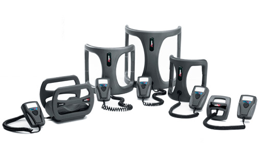
What treatment options are there for non-union fractures?
There are two types of treatment options that can be utilized for non-union fractures
-
- Non-Surgical: Bone growth stimulators that include electrical stimulation, ultrasound or shock wave therapy.
- Surgical: There are numerous operative techniques for non-union fractures, the most common are external fixation with hardware, plating, or bone grafting.
[landing_block type=”cta_buttons”]
References
- Behrens SB, Deren ME, Monchik KO. A review of bone growth stimulation for fracture treatment. Curr Orthop Pract. 2013; 24(1):84-91.
- Rabjohn, Linnie V. DPM. Electrical Stimulation of Bone: The Evolving Technology. Clinical Podiatry, Podiatry Management. 2008; 167 -176.
- Summers, Hobie MD, Daniel S. Chan MD. General Principles in the Assessment and Treatment of Nonunions. April 2011